Section 49-4 Review Drugs and the Nervous System Answers
| Structure of a typical chemical synapse |
|---|
| Postsynaptic Voltage- Synaptic Neurotransmitter Receptor Neurotransmitter Axon terminal Synaptic cleft Dendrite |
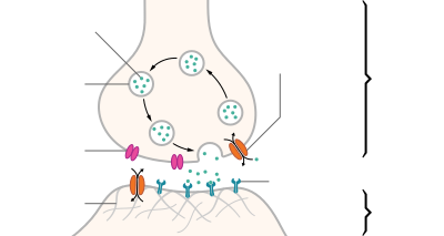
A neurotransmitter is a signaling molecule secreted by a neuron to affect another cell across a synapse. The jail cell receiving the signal, any main trunk part or target cell, may be another neuron, only could also be a gland or muscle cell.[1]
Neurotransmitters are released from synaptic vesicles into the synaptic cleft where they are able to collaborate with neurotransmitter receptors on the target cell. The neurotransmitter's event on the target prison cell is determined by the receptor information technology binds. Many neurotransmitters are synthesized from simple and plentiful precursors such every bit amino acids, which are readily available and frequently crave a small number of biosynthetic steps for conversion.
Neurotransmitters are essential to the role of complex neural systems. The verbal number of unique neurotransmitters in humans is unknown, but more than 100 have been identified.[2] Common neurotransmitters include glutamate, GABA, acetylcholine, glycine and norepinephrine.
Mechanism and bike [edit]
Synthesis [edit]
Neurotransmitters are more often than not synthesized in neurons and are made up of, or derived from, forerunner molecules that are constitute abundantly in the cell. Classes of neurotransmitters include amino acids, monoamines, and peptides. Monoamines are synthesized by altering a unmarried amino acid. For case, the precursor of serotonin is the amino acid tryptophan. Peptide transmitters, or neuropeptides, are protein transmitters that often are released together with other transmitters to have a modulatory effect.[iii] Purine neurotransmitters, similar ATP, are derived from nucleic acids. Other neurotransmitters are made up of metabolic products similar nitric oxide and carbon monoxide.
| Examples | |
|---|---|
| Amino Acrid | glycine, glutamate |
| Monoamines | serotonin, epinephrine, dopamine |
| Peptides | substance P, opioids |
| Purines | ATP, GTP |
| Other | nitric oxide, carbon monoxide |
![]()
Synaptic vesicles containing neurotransmitters
Storage [edit]
Neurotransmitters are generally stored in synaptic vesicles, clustered shut to the jail cell membrane at the axon terminal of the presynaptic neuron. Still, some neurotransmitters, similar the metabolic gases carbon monoxide and nitric oxide, are synthesized and released immediately post-obit an action potential without always being stored in vesicles.[4]
Release [edit]
Generally, a neurotransmitter is released at the presynaptic terminal in response to an electric bespeak called an activity potential in the presynaptic neuron. However, low level 'baseline' release also occurs without electrical stimulation. Neurotransmitters are released into and diffuse beyond the synaptic cleft, where they demark to specific receptors on the membrane of the postsynaptic neuron.[five]
Receptor interaction [edit]
Subsequently beingness released into the synaptic fissure, neurotransmitters diffuse across the synapse where they are able to interact with receptors on the target cell. The event of the neurotransmitter is dependent on the identity of the target cell's receptors present at the synapse. Depending on the receptor, binding of neurotransmitters may crusade excitation, inhibition, or modulation of the postsynaptic neuron. See below for more information.
Elimination [edit]
Acetylcholine is cleaved in the synaptic scissure into acerb acid and choline
In order to avoid continuous activation of receptors on the post-synaptic or target cell, neurotransmitters must be removed from the synaptic cleft.[half dozen] Neurotransmitters are removed through one of 3 mechanisms:
- Diffusion – neurotransmitters drift out of the synaptic cleft, where they are absorbed by glial cells. These glial cells, usually astrocytes, absorb the excess neurotransmitters. In the glial jail cell, neurotransmitters are cleaved down by enzymes or pumped dorsum into
- Enzyme degradation – proteins chosen enzymes break the neurotransmitters down.
- Reuptake – neurotransmitters are reabsorbed into the pre-synaptic neuron. Transporters, or membrane ship proteins, pump neurotransmitters from the synaptic cleft back into axon terminals (the presynaptic neuron) where they are stored for reuse.
For example, acetylcholine is eliminated past having its acetyl group broken by the enzyme acetylcholinesterase; the remaining choline is and so taken in and recycled by the pre-synaptic neuron to synthesize more acetylcholine.[7] Other neurotransmitters are able to diffuse away from their targeted synaptic junctions and are eliminated from the body via the kidneys, or destroyed in the liver. Each neurotransmitter has very specific degradation pathways at regulatory points, which may be targeted by the body'south regulatory organisation or medication. Cocaine blocks a dopamine transporter responsible for the reuptake of dopamine. Without the transporter, dopamine diffuses much more than slowly from the synaptic cleft and continues to actuate the dopamine receptors on the target cell.[8]
Discovery [edit]
Until the early 20th century, scientists causeless that the majority of synaptic communication in the brain was electrical. However, through histological examinations by Ramón y Cajal, a 20 to xl nm gap between neurons, known today equally the synaptic cleft, was discovered. The presence of such a gap suggested communication via chemical messengers traversing the synaptic crevice, and in 1921 German pharmacologist Otto Loewi confirmed that neurons can communicate by releasing chemicals. Through a series of experiments involving the vagus fretfulness of frogs, Loewi was able to manually slow the heart charge per unit of frogs by controlling the corporeality of saline solution present effectually the vagus nervus. Upon completion of this experiment, Loewi asserted that sympathetic regulation of cardiac function can be mediated through changes in chemic concentrations. Furthermore, Otto Loewi is credited with discovering acetylcholine (ACh) – the beginning known neurotransmitter.[ix]
Identification [edit]
There are four master criteria for identifying neurotransmitters:
- The chemical must be synthesized in the neuron or otherwise be present in it.
- When the neuron is active, the chemical must be released and produce a response in some targets.
- The same response must be obtained when the chemical is experimentally placed on the target.
- A machinery must exist for removing the chemical from its site of activation after its work is washed.
Nonetheless, given advances in pharmacology, genetics, and chemical neuroanatomy, the term "neurotransmitter" tin be practical to chemicals that:
- Bear letters between neurons via influence on the postsynaptic membrane.
- Have little or no upshot on membrane voltage, just take a common conveying function such every bit changing the construction of the synapse.
- Communicate by sending reverse-direction messages that affect the release or reuptake of transmitters.
The anatomical localization of neurotransmitters is typically determined using immunocytochemical techniques, which place the location of either the transmitter substances themselves or of the enzymes that are involved in their synthesis. Immunocytochemical techniques take also revealed that many transmitters, peculiarly the neuropeptides, are co-localized, that is, a neuron may release more than one transmitter from its synaptic final.[x] Various techniques and experiments such as staining, stimulating, and collecting tin be used to identify neurotransmitters throughout the primal nervous organization.[11]
Deportment [edit]
Neurons form elaborate networks through which nerve impulses – activity potentials – travel. Each neuron has every bit many every bit xv,000 connections with neighboring neurons.
Neurons do not touch each other (except in the case of an electrical synapse through a gap junction); instead, neurons interact at contact points called synapses: a junction within ii nerve cells, consisting of a miniature gap within which impulses are carried by a neurotransmitter. A neuron transports its information past way of a nerve impulse chosen an action potential. When an activeness potential arrives at the synapse's presynaptic terminal push, it may stimulate the release of neurotransmitters. These neurotransmitters are released into the synaptic cleft to demark onto the receptors of the postsynaptic membrane and influence some other cell, either in an inhibitory or excitatory style. The next neuron may be connected to many more neurons, and if the total of excitatory influences minus inhibitory influences is great enough, it will likewise "fire". That is to say, it will create a new activeness potential at its axon hillock, releasing neurotransmitters and passing on the information to yet another neighboring neuron.
Modulation [edit]
A neurotransmitter may have an excitatory, inhibitory or modulatory effect on the target cell. The effect is determined past the receptors the neurotransmitter interacts with at the postal service-synaptic membrane. Neurotransmitter influences trans-membrane ion menstruation either to increase (excitatory) or to subtract (inhibitory) the probability that the cell with which it comes in contact will produce an activeness potential. Synapses containing receptors with excitatory effects are called Blazon I synapses, while Type 2 synapses contain receptors with inhibitory furnishings.[12] Thus, despite the wide variety of synapses, they all convey messages of only these two types. The two types are different advent and are primarily located on unlike parts of the neurons under its influence.[13] Receptors with modulatory effects are spread throughout all synaptic membranes and binding of neurotransmitters sets in motion signaling cascades that assistance the cell regulate its part.[14] Binding of neurotransmitters to receptors with modulatory effects can have many results. For example it may result in an increase or decrease in sensitivity to future stimulus by recruiting more or less receptors to the synaptic membrane.
Type I (excitatory) synapses are typically located on the shafts or the spines of dendrites, whereas type II (inhibitory) synapses are typically located on a cell body. In improver, Type I synapses have round synaptic vesicles, whereas the vesicles of type II synapses are flattened. The material on the presynaptic and post-synaptic membranes is denser in a Type I synapse than it is in a type II, and the type I synaptic cleft is wider. Finally, the active zone on a Type I synapse is larger than that on a Blazon II synapse.
The unlike locations of type I and blazon II synapses split up a neuron into two zones: an excitatory dendritic tree and an inhibitory prison cell torso. From an inhibitory perspective, excitation comes in over the dendrites and spreads to the axon hillock to trigger an action potential. If the message is to be stopped, it is best stopped by applying inhibition on the cell body, close to the axon hillock where the activity potential originates. Another fashion to anticipate excitatory–inhibitory interaction is to moving-picture show excitation overcoming inhibition. If the cell trunk is ordinarily in an inhibited land, the just way to generate an action potential at the axon hillock is to reduce the jail cell body's inhibition. In this "open the gates" strategy, the excitatory bulletin is like a racehorse ready to run downward the rails, but offset, the inhibitory starting gate must be removed.[15]
Neurotransmitter deportment [edit]
As explained above, the only direct activity of a neurotransmitter is to actuate a receptor. Therefore, the effects of a neurotransmitter system depend on the connections of the neurons that use the transmitter, and the chemical properties of the receptors.
- Glutamate is used at the great majority of fast excitatory synapses in the brain and spinal cord. Information technology is also used at almost synapses that are "modifiable", i.e. capable of increasing or decreasing in strength. Modifiable synapses are thought to be the main retentivity-storage elements in the brain. Excessive glutamate release can overstimulate the brain and lead to excitotoxicity causing cell decease resulting in seizures or strokes.[16] Excitotoxicity has been implicated in certain chronic diseases including ischemic stroke, epilepsy, amyotrophic lateral sclerosis, Alzheimer's disease, Huntington affliction, and Parkinson's affliction.[17]
- GABA is used at the great majority of fast inhibitory synapses in virtually every part of the brain. Many allaying/tranquilizing drugs human activity by enhancing the effects of GABA.[18] Correspondingly, glycine is the inhibitory transmitter in the spinal string.
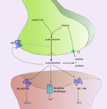
- Acetylcholine was the first neurotransmitter discovered in the peripheral and central nervous systems. Information technology activates skeletal muscles in the somatic nervous arrangement and may either excite or inhibit internal organs in the autonomic arrangement.[xi] It is distinguished as the transmitter at the neuromuscular junction connecting motor nerves to muscles. The paralytic arrow-poisonous substance curare acts by blocking manual at these synapses. Acetylcholine besides operates in many regions of the brain, but using different types of receptors, including nicotinic and muscarinic receptors.[19]
- Dopamine has a number of important functions in the encephalon; this includes regulation of motor behavior, pleasures related to motivation and too emotional arousal. It plays a critical role in the advantage organisation; Parkinson'south illness has been linked to low levels of dopamine and schizophrenia has been linked to loftier levels of dopamine.[20]
- Serotonin is a monoamine neurotransmitter. Well-nigh is produced by and found in the intestine (approximately ninety%), and the remainder in central nervous system neurons. It functions to regulate appetite, sleep, memory and learning, temperature, mood, behaviour, muscle contraction, and function of the cardiovascular system and endocrine organisation. It is speculated to have a part in depression, as some depressed patients are seen to have lower concentrations of metabolites of serotonin in their cerebrospinal fluid and brain tissue.[21]
- Norepinephrine which is synthesized in the central nervous system and sympathetic fretfulness, modulates the responses of the autonomic nervous arrangement, the slumber patterns, focus and alacrity. It is synthesized from tyrosine.
- Epinephrine which is also synthesized from tyrosine is released in the adrenal glands and the brainstem. It plays a role in sleep, with one's power to get and stay alert, and the fight-or-flight response.
Types [edit]
At that place are many different means to classify neurotransmitters. Dividing them into amino acids, peptides, and monoamines is sufficient for some classification purposes.[22]
Major neurotransmitters:
- Amino acids: glutamate,[23] aspartate, D-serine, gamma-Aminobutyric acid (GABA),[nb 1] glycine
- Gasotransmitters: nitric oxide (NO), carbon monoxide (CO), hydrogen sulfide (HiiS)
- Monoamines: dopamine (DA), norepinephrine (noradrenaline; NE, NA), epinephrine (adrenaline), histamine, serotonin (SER, 5-HT)
- Catecholamines: dopamine, norepinephrine (noradrenaline), epinephrine (adrenaline)
- Trace amines: phenethylamine, N-methylphenethylamine, tyramine, iii-iodothyronamine, octopamine, tryptamine, etc.
- Peptides: oxytocin, somatostatin, substance P, cocaine and amphetamine regulated transcript, opioid peptides[24]
- Purines: adenosine triphosphate (ATP), adenosine
- Others: acetylcholine (ACh), anandamide, etc.
In addition, over 100 neuroactive peptides accept been found, and new ones are discovered regularly.[25] [26] Many of these are co-released along with a small-molecule transmitter. All the same, in some cases, a peptide is the main transmitter at a synapse. Beta-Endorphin is a relatively well-known example of a peptide neurotransmitter because it engages in highly specific interactions with opioid receptors in the central nervous system.
Single ions (such as synaptically released zinc) are also considered neurotransmitters past some,[27] as well as some gaseous molecules such as nitric oxide (NO), carbon monoxide (CO), and hydrogen sulfide (H2S).[28] The gases are produced in the neural cytoplasm and are immediately diffused through the cell membrane into the extracellular fluid and into nearby cells to stimulate product of 2d messengers. Soluble gas neurotransmitters are difficult to study, as they act rapidly and are immediately broken downwards, existing for only a few seconds.
The most prevalent transmitter is glutamate, which is excitatory at well over 90% of the synapses in the human encephalon.[23] The adjacent most prevalent is gamma-Aminobutyric Acid, or GABA, which is inhibitory at more than 90% of the synapses that do not use glutamate. Although other transmitters are used in fewer synapses, they may be very important functionally: the corking majority of psychoactive drugs exert their effects by altering the actions of some neurotransmitter systems, oft acting through transmitters other than glutamate or GABA. Addictive drugs such equally cocaine and amphetamines exert their furnishings primarily on the dopamine organization. The addictive opiate drugs exert their effects primarily equally functional analogs of opioid peptides, which, in turn, regulate dopamine levels.
List of neurotransmitters, peptides, and gaseous signaling molecules [edit]
| Category | Name | Abbreviation | Metabotropic | Ionotropic |
|---|---|---|---|---|
| Minor: Amino acids (Arg) | Arginine | Arg, R | α2-Adrenergic receptors, imidazoline receptors | NMDA receptors |
| Small-scale: Amino acids | Aspartate | Asp, D | – | NMDA receptors |
| Small: Amino acids | Glutamate | Glu, E | Metabotropic glutamate receptors | NMDA receptors, kainate receptors, AMPARs |
| Pocket-sized: Amino acids | Gamma-aminobutyric acid | GABA | GABAB receptors | GABAA receptors, GABAA-ρ receptors |
| Pocket-size: Amino acids | Glycine | Gly, One thousand | – | NMDA receptors, glycine receptors |
| Small: Amino acids | D-serine | Ser, Southward | – | NMDA receptors |
| Small: Acetylcholine | Acetylcholine | ACh | Muscarinic acetylcholine receptors | Nicotinic acetylcholine receptors |
| Small: Monoamine (Phe/Tyr) | Dopamine | DA | Dopamine receptors, trace amine-associated receptor one[29] [xxx] | – |
| Small-scale: Monoamine (Phe/Tyr) | Norepinephrine (noradrenaline) | NE, NAd | Adrenergic receptors | – |
| Small: Monoamine (Phe/Tyr) | Epinephrine (adrenaline) | Epi, Advert | Adrenergic receptors | – |
| Small: Monoamine (Trp) | Serotonin (five-hydroxytryptamine) | 5-HT | Serotonin receptors (all except 5-HT3) | v-HT3 |
| Small: Monoamine (His) | Histamine | H | Histamine receptors | – |
| Pocket-sized: Trace amine (Phe) | Phenethylamine | PEA | Homo trace amine-associated receptors: hTAAR1, hTAAR2 | – |
| Small-scale: Trace amine (Phe) | Northward-methylphenethylamine | NMPEA | hTAAR1 | – |
| Small-scale: Trace amine (Phe/Tyr) | Tyramine | TYR | hTAAR1, hTAAR2 | – |
| Small: Trace amine (Phe/Tyr) | octopamine | Oct | hTAAR1 | – |
| Small: Trace amine (Phe/Tyr) | Synephrine | Syn | hTAAR1 | – |
| Small: Trace amine (Trp) | Tryptamine | hTAAR1, various serotonin receptors | – | |
| Small: Trace amine (Trp) | N-methyltryptamine | NMT | hTAAR1, various serotonin receptors | – |
| Lipid | Anandamide | AEA | Cannabinoid receptors | – |
| Lipid | 2-Arachidonoylglycerol | 2-AG | Cannabinoid receptors | – |
| Lipid | ii-Arachidonyl glyceryl ether | 2-AGE | Cannabinoid receptors | – |
| Lipid | Due north-Arachidonoyl dopamine | Goose egg | Cannabinoid receptors | TRPV1 |
| Lipid | Virodhamine | Cannabinoid receptors | – | |
| Small: Purine | Adenosine | Ado | Adenosine receptors | – |
| Small: Purine | Adenosine triphosphate | ATP | P2Y receptors | P2X receptors |
| Pocket-sized: Purine | Nicotinamide adenine dinucleotide | β-NAD | P2Y receptors | P2X receptors |
| Category | Proper noun | Abbreviation | Metabotropic | Ionotropic |
|---|---|---|---|---|
| Bombesin-like peptides | Bombesin | BBR1-2-3 | – | |
| Bombesin-like peptide | Gastrin releasing peptide | GRP | – | – |
| Bombesin-similar peptide | Neuromedin B | NMB | Neuromedin B receptor | – |
| Bradykinins | Bradykinin | B1, B2 | – | |
| Calcitonin/CGRP family | Calcitonin | Calcitonin receptor | – | |
| Calcitonin/CGRP family unit | Calcitonin cistron-related peptide | CGRP | CALCRL | – |
| Corticotropin-releasing factors | Corticotropin-releasing hormone | CRH | CRHR1 | – |
| Corticotropin-releasing factors | Urocortin | CRHR1 | – | |
| Galanins | Galanin | GALR1, GALR2, GALR3 | – | |
| Galanins | Galanin-like peptide | GALR1, GALR2, GALR3 | – | |
| Gastrins | Gastrin | Cholecystokinin B receptor | – | |
| Gastrins | Cholecystokinin | CCK | Cholecystokinin receptors | – |
| Granins | Chromogranin A | ChgA | – | – |
| Melanocortins | Adrenocorticotropic hormone | ACTH | ACTH receptor | – |
| Melanocortins | Proopiomelanocortin | POMC | Melanocortin four receptor | – |
| Melanocortins | Melanocyte-stimulating hormones | MSH | Melanocortin receptors | – |
| Neurohypophyseals | Vasopressin | AVP | Vasopressin receptors | – |
| Neurohypophyseals | Oxytocin | OT | Oxytocin receptor | – |
| Neurohypophyseals | Neurophysin I | – | – | |
| Neurohypophyseals | Neurophysin Ii | – | – | |
| Neurohypophyseals | Copeptin | – | – | |
| Neuromedins | Neuromedin U | NmU | NmUR1, NmUR2 | – |
| Neuropeptide B/W | Neuropeptide B | NPB | NPBW1, NPBW2 | – |
| Neuropeptide B/W | Neuropeptide South | NPS | Neuropeptide Due south receptors | – |
| Neuropeptide Y | Neuropeptide Y | NY | Neuropeptide Y receptors | – |
| Neuropeptide Y | Pancreatic polypeptide | PP | – | – |
| Neuropeptide Y | Peptide YY | PYY | – | – |
| Opioids | Enkephalins | δ-Opioid receptor | – | |
| Opioids | Dynorphins | κ-Opioid receptor | – | |
| Opioids | Neoendorphins | κ-Opioid receptor | – | |
| Opioids | Endorphins | μ-Opioid receptors | – | |
| Opioids | Endomorphins | μ-Opioid receptors | – | |
| Opioids | Morphine | μ-Opioid receptors | – | |
| Opioids | Nociceptin/orphanin FQ | Due north/OFQ | Nociceptin receptors | – |
| Orexins | Orexin A | OX-A | Orexin receptors | – |
| Orexins | Orexin B | OX-B | Orexin receptors | – |
| Parathyroid hormone family | Parathyroid hormone-related protein | PTHrP | – | – |
| RFamides | Kisspeptin | Osculation | GPR54 | – |
| RFamides | Neuropeptide FF | NPFF | NPFF1, NPFF2 | – |
| RFamides | Prolactin-releasing peptide | PrRP | PrRPR | – |
| RFamides | Pyroglutamylated RFamide peptide | QRFP | GPR103 | – |
| Secretins | Secretin | Secretin receptor | – | |
| Secretins | Motilin | Motilin receptor | – | |
| Secretins | Glucagon | Glucagon receptor | – | |
| Secretins | Glucagon-like peptide-1 | GLP-one | Glucagon-like peptide one receptor | – |
| Secretins | Glucagon-like peptide-two | GLP-2 | Glucagon-like peptide 2 receptor | – |
| Secretins | Vasoactive intestinal peptide | VIP | Vasoactive intestinal peptide receptors | – |
| Secretins | Growth hormone–releasing hormone | GHRH | Growth hormone–releasing hormone receptor | – |
| Secretins | Pituitary adenylate cyclase-activating peptide | PACAP | ADCYAP1R1 | – |
| Somatostatins | Somatostatin | Somatostatin receptors | – | |
| Tachykinins | Neurokinin A | – | – | |
| Tachykinins | Neurokinin B | – | – | |
| Tachykinins | Substance P | – | – | |
| Tachykinins | Neuropeptide K | – | – | |
| Other | Agouti-related peptide | AgRP | Melanocortin receptor – | |
| Other | N-Acetylaspartylglutamate | NAAG | Metabotropic glutamate receptor iii (mGluR3) | – |
| Other | Cocaine- and amphetamine-regulated transcript | CART | Unknown One thousandi/One thousando-coupled receptor[31] | – |
| Other | Gonadotropin-releasing hormone | GnRH | GnRHR | – |
| Other | Thyrotropin-releasing hormone | TRH | TRHR | – |
| Other | Melanin-concentrating hormone | MCH | MCHR 1,2 | – |
| Category | Name | Abbreviation | Metabotropic | Ionotropic |
|---|---|---|---|---|
| Gaseous signaling molecule | Nitric oxide | NO | Soluble guanylyl cyclase | – |
| Gaseous signaling molecule | Carbon monoxide | CO | – | Heme jump to potassium channels |
| Gaseous signaling molecule | Hydrogen sulfide | H2S | – | – |
Encephalon neurotransmitter systems [edit]
Neurons expressing certain types of neurotransmitters sometimes form singled-out systems, where activation of the system affects large volumes of the brain, chosen volume transmission. Major neurotransmitter systems include the noradrenaline (norepinephrine) system, the dopamine system, the serotonin system, and the cholinergic organization, amongst others. Trace amines accept a modulatory effect on neurotransmission in monoamine pathways (i.e., dopamine, norepinephrine, and serotonin pathways) throughout the encephalon via signaling through trace amine-associated receptor 1.[32] [33] A brief comparison of these systems follows:
| System | Pathway origin and projections | Regulated cognitive processes and behaviors |
|---|---|---|
| Noradrenaline arrangement [34] [35] [36] [37] [38] [39] | Noradrenergic pathways:
|
|
| Dopamine system [36] [37] [38] [40] [41] [42] | Dopaminergic pathways:
|
|
| Histamine system [37] [38] [43] | Histaminergic pathways:
|
|
| Serotonin system [34] [36] [37] [38] [44] [45] [46] | Serotonergic pathways: Caudal nuclei (CN):
Rostral nuclei (RN):
|
|
| Acetylcholine system [34] [36] [37] [38] [47] | Cholinergic pathways: Forebrain cholinergic nuclei (FCN):
Striatal tonically active cholinergic neurons (TAN)
Brainstem cholinergic nuclei (BCN):
|
|
| Adrenaline system [48] [49] | Adrenergic pathways:
|
|
Drug effects [edit]
Understanding the furnishings of drugs on neurotransmitters comprises a significant portion of research initiatives in the field of neuroscience. Most neuroscientists involved in this field of research believe that such efforts may further advance our agreement of the circuits responsible for various neurological diseases and disorders, as well equally ways to effectively treat and someday possibly prevent or cure such illnesses.[fifty] [ medical citation needed ]
Drugs can influence behavior by altering neurotransmitter action. For example, drugs tin can decrease the charge per unit of synthesis of neurotransmitters by affecting the synthetic enzyme(s) for that neurotransmitter. When neurotransmitter syntheses are blocked, the amount of neurotransmitters available for release becomes substantially lower, resulting in a decrease in neurotransmitter activeness. Some drugs block or stimulate the release of specific neurotransmitters. Alternatively, drugs tin can prevent neurotransmitter storage in synaptic vesicles past causing the synaptic vesicle membranes to leak. Drugs that prevent a neurotransmitter from binding to its receptor are called receptor antagonists. For example, drugs used to treat patients with schizophrenia such as haloperidol, chlorpromazine, and clozapine are antagonists at receptors in the brain for dopamine. Other drugs act past binding to a receptor and mimicking the normal neurotransmitter. Such drugs are called receptor agonists. An example of a receptor agonist is morphine, an opiate that mimics effects of the endogenous neurotransmitter β-endorphin to relieve hurting. Other drugs interfere with the deactivation of a neurotransmitter after it has been released, thereby prolonging the action of a neurotransmitter. This can be accomplished past blocking re-uptake or inhibiting degradative enzymes. Lastly, drugs can also prevent an action potential from occurring, blocking neuronal activity throughout the central and peripheral nervous system. Drugs such as tetrodotoxin that block neural activity are typically lethal.
Drugs targeting the neurotransmitter of major systems affect the whole arrangement, which can explain the complication of action of some drugs. Cocaine, for example, blocks the re-uptake of dopamine back into the presynaptic neuron, leaving the neurotransmitter molecules in the synaptic gap for an extended period of time. Since the dopamine remains in the synapse longer, the neurotransmitter continues to demark to the receptors on the postsynaptic neuron, eliciting a pleasurable emotional response. Concrete addiction to cocaine may consequence from prolonged exposure to excess dopamine in the synapses, which leads to the downregulation of some post-synaptic receptors. Subsequently the effects of the drug wear off, an individual can become depressed due to decreased probability of the neurotransmitter bounden to a receptor. Fluoxetine is a selective serotonin re-uptake inhibitor (SSRI), which blocks re-uptake of serotonin by the presynaptic jail cell which increases the amount of serotonin present at the synapse and furthermore allows information technology to remain there longer, providing potential for the effect of naturally released serotonin.[51] AMPT prevents the conversion of tyrosine to L-DOPA, the precursor to dopamine; reserpine prevents dopamine storage within vesicles; and deprenyl inhibits monoamine oxidase (MAO)-B and thus increases dopamine levels.
| Drug | Interacts with: | Receptor Interaction: | Type | Effects |
|---|---|---|---|---|
| Botulinum Toxin (Botox) | Acetylcholine | – | Adversary | Blocks acetylcholine release in PNS Prevents muscle contractions |
| Black Widow Spider Venom | Acetylcholine | – | Agonist | Promotes acetylcholine release in PNS Stimulates musculus contractions |
| Neostigmine | Acetylcholine | – | – | Interferes with acetylcholinerase activity Increases effects of ACh at receptors Used to treat myasthenia gravis |
| Nicotine | Acetylcholine | Nicotinic (skeletal muscle) | Agonist | Increases ACh activity Increases attending Reinforcing effects |
| d-tubocurarine | Acetylcholine | Nicotinic (skeletal musculus) | Antagonist | Decreases activity at receptor site |
| Curare | Acetylcholine | Nicotinic (skeletal muscle) | Antagonist | Decreases ACh activity Prevents muscle contractions |
| Muscarine | Acetylcholine | Muscarinic (heart and smooth muscle) | Agonist | Increases ACh action Toxic |
| Atropine | Acetylcholine | Muscarinic (heart and smooth muscle) | Adversary | Blocks educatee constriction Blocks saliva production |
| Scopolamine (Hyoscine) | Acetylcholine | Muscarinic (heart and smoothen muscle) | Antagonist | Treats motion sickness and postoperative nausea and vomiting |
| AMPT | Dopamine/norepinephrine | – | – | Inactivates tyrosine hydroxylase and inhibits dopamine production |
| Reserpine | Dopamine | – | – | Prevents storage of dopamine and other monoamines in synaptic vesicles Causes sedation and depression |
| Apomorphine | Dopamine | D2 Receptor (presynaptic autoreceptors/postsynaptic receptors) | Antagonist (depression dose)/Straight agonist (high dose) | Low dose: blocks autoreceptors High dose: stimulates postsynaptic receptors |
| Amphetamine | Dopamine/norepinephrine | – | Indirect agonist | Releases dopamine, noradrenaline, and serotonin Blocks reuptake[32] [33] |
| Methamphetamine | Dopamine/norepinephrine | – | – | Releases dopamine and noradrenaline Blocks reuptake |
| Methylphenidate | Dopamine | – | – | Blocks reuptake Enhances attention and impulse control in ADHD |
| Cocaine | Dopamine | – | Indirect Agonist | Blocks reuptake into presynapse Blocks voltage-dependent sodium channels Tin can be used every bit a topical anesthetic (eye drops) |
| Deprenyl | Dopamine | – | Agonist | Inhibits MAO-B Prevents destruction of dopamine |
| Chlorpromazine | Dopamine | D2 Receptors | Antagonist | Blocks D2 receptors Alleviates hallucinations |
| MPTP | Dopamine | – | – | Results in Parkinson like symptoms |
| PCPA | Serotonin (5-HT) | – | Antagonist | Disrupts serotonin synthesis by blocking the activity of tryptophan hydroxylase |
| Ondansetron | Serotonin (5-HT) | v-HTthree receptors | Antagonist | Reduces side effects of chemotherapy and radiation Reduces nausea and airsickness |
| Buspirone | Serotonin (5-HT) | 5-HT1A receptors | Partial Agonist | Treats symptoms of anxiety and depression |
| Fluoxetine | Serotonin (5-HT) | supports 5-HT reuptake | SSRI | Inhibits reuptake of serotonin Treats low, some anxiety disorders, and OCD[51] Mutual examples: Prozac and Sarafem |
| Fenfluramine | Serotonin (v-HT) | – | – | Causes release of serotonin Inhibits reuptake of serotonin Used equally an appetite suppressant |
| Lysergic acrid diethylamide | Serotonin (5-HT) | Post-synaptic 5-HT2A receptors | Direct Agonist | Produces visual perception distortions Stimulates five-HT2A receptors in forebrain |
| Methylenedioxymethamphetamine (MDMA) | Serotonin (5-HT)/ norepinphrine | – | – | Stimulates release of serotonin and norepinephrine and inhibits the reuptake Causes excitatory and hallucinogenic furnishings |
| Strychnine | Glycine | – | Antagonist | Causes severe muscle spasms[53] |
| Diphenhydramine | Histamine | Crosses blood encephalon bulwark to cause drowsiness | ||
| Tetrahydrocannabinol (THC) | Endocannabinoids | Cannabinoid (CB) receptors | Agonist | Produces analgesia and sedation Increases appetite Cognitive effects |
| Rimonabant | Endocannabinoids | Cannabinoid (CB) receptors | Antagonist | Suppresses appetite Used in smoking cessation |
| MAFP | Endocannabinoids | – | – | Inhibits FAAH Used in enquiry to increase cannabinoid organisation activity |
| AM1172 | Endocannabinoids | – | – | Blocks cannabinoid reuptake Used in research to increase cannabinoid system activity |
| Anandamide (endogenous) | – | Cannabinoid (CB) receptors; 5-HTthree receptors | – | Reduce nausea and vomiting |
| Caffeine | Adenosine | Adenosine receptors | Antagonist | Blocks adenosine receptors Increases wakefulness |
| PCP | Glutamate | NMDA receptor | Indirect Antagonist | Blocks PCP bounden site Prevents calcium ions from entering neurons Impairs learning |
| AP5 | Glutamate | NMDA receptor | Antagonist | Blocks glutamate bounden site on NMDA receptor Impairs synaptic plasticity and certain forms of learning |
| Ketamine | Glutamate | NMDA receptor | Adversary | Used as anesthesia Induces trance-similar state, helps with hurting relief and sedation |
| NMDA | Glutamate | NMDA receptor | Agonist | Used in research to written report NMDA receptor Ionotropic receptor |
| AMPA | Glutamate | AMPA receptor | Agonist | Used in research to written report AMPA receptor Ionotropic receptor |
| Allyglycine | GABA | – | – | Inhibits GABA synthesis Causes seizures |
| Muscimol | GABA | GABA receptor | Agonist | Causes sedation |
| Bicuculine | GABA | GABA receptor | Adversary | Causes Seizures |
| Benzodiazepines | GABA | GABAA receptor | Indirect agonists | Anxiolytic, sedation, memory impairment, muscle relaxation |
| Barbiturates | GABA | GABAA receptor | Indirect agonists | Sedation, retention harm, muscle relaxation |
| Booze | GABA | GABA receptor | Indirect agonist | Sedation, retentiveness damage, musculus relaxation |
| Picrotoxin | GABA | GABAA receptor | Indirect adversary | High doses cause seizures |
| Tiagabine | GABA | – | Antagonist | GABA transporter antagonist Increment availability of GABA Reduces the likelihood of seizures |
| Moclobemide | Norepinephrine | – | Agonist | Blocks MAO-A to treat depression |
| Idazoxan | Norepinephrine | alpha-ii adrenergic autoreceptors | Agonist | Blocks blastoff-2 autoreceptors Used to study norepinephrine organisation |
| Fusaric acrid | Norepinephrine | – | – | Inhibits activeness of dopamine beta-hydroxylase which blocks the product of norepinephrine Used to written report norepinephrine system without affecting dopamine system |
| Opiates (Opium, morphine, heroin, and oxycodone) | Opioids | Opioid receptor[54] | Agonists | Analgesia, sedation, and reinforcing furnishings |
| Naloxone | Opioids | – | Adversary | Reverses opiate intoxication or overdose symptoms (i.east. bug with breathing) |
Agonists [edit]
| | This section needs expansion with: coverage of total agonists and their distinction from partial agonist and inverse agonist.. You lot can help by adding to it. (August 2015) |
An agonist is a chemical capable of bounden to a receptor, such every bit a neurotransmitter receptor, and initiating the same reaction typically produced by the bounden of the endogenous substance.[55] An agonist of a neurotransmitter volition thus initiate the same receptor response as the transmitter. In neurons, an agonist drug may activate neurotransmitter receptors either directly or indirectly. Direct-binding agonists can be farther characterized as full agonists, partial agonists, inverse agonists.[56] [57]
Straight agonists act similar to a neurotransmitter by binding straight to its associated receptor site(south), which may be located on the presynaptic neuron or postsynaptic neuron, or both.[58] Typically, neurotransmitter receptors are located on the postsynaptic neuron, while neurotransmitter autoreceptors are located on the presynaptic neuron, as is the case for monoamine neurotransmitters;[32] in some cases, a neurotransmitter utilizes retrograde neurotransmission, a type of feedback signaling in neurons where the neurotransmitter is released postsynaptically and binds to target receptors located on the presynaptic neuron.[59] [annotation 1] Nicotine, a compound found in tobacco, is a straight agonist of most nicotinic acetylcholine receptors, mainly located in cholinergic neurons.[54] Opiates, such every bit morphine, heroin, hydrocodone, oxycodone, codeine, and methadone, are μ-opioid receptor agonists; this action mediates their euphoriant and pain relieving properties.[54]
Indirect agonists increase the binding of neurotransmitters at their target receptors by stimulating the release or preventing the reuptake of neurotransmitters.[58] Some indirect agonists trigger neurotransmitter release and prevent neurotransmitter reuptake. Amphetamine, for example, is an indirect agonist of postsynaptic dopamine, norepinephrine, and serotonin receptors in each their respective neurons;[32] [33] it produces both neurotransmitter release into the presynaptic neuron and after the synaptic scissure and prevents their reuptake from the synaptic fissure by activating TAAR1, a presynaptic K protein-coupled receptor, and binding to a site on VMAT2, a blazon of monoamine transporter located on synaptic vesicles inside monoamine neurons.[32] [33]
Antagonists [edit]
An antagonist is a chemical that acts within the torso to reduce the physiological activity of another chemic substance (as an opiate); peculiarly one that opposes the action on the nervous system of a drug or a substance occurring naturally in the trunk by combining with and blocking its nervous receptor.[lx]
In that location are two primary types of antagonist: direct-acting Antagonist and indirect-interim Antagonists:
- Direct-acting antagonist- which takes up space nowadays on receptors which are otherwise taken up past neurotransmitters themselves. This results in neurotransmitters existence blocked from binding to the receptors. The well-nigh common is chosen Atropine.
- Indirect-acting antagonist- drugs that inhibit the release/production of neurotransmitters (e.thou., Reserpine).
Drug antagonists [edit]
An antagonist drug is one that attaches (or binds) to a site chosen a receptor without activating that receptor to produce a biological response. It is therefore said to have no intrinsic activity. An adversary may also exist called a receptor "blocker" because they cake the outcome of an agonist at the site. The pharmacological furnishings of an antagonist, therefore, upshot in preventing the corresponding receptor site's agonists (e.1000., drugs, hormones, neurotransmitters) from bounden to and activating information technology. Antagonists may exist "competitive" or "irreversible".
A competitive antagonist competes with an agonist for binding to the receptor. Equally the concentration of adversary increases, the binding of the agonist is progressively inhibited, resulting in a subtract in the physiological response. Loftier concentration of an adversary can completely inhibit the response. This inhibition can exist reversed, however, by an increment of the concentration of the agonist, since the agonist and antagonist compete for binding to the receptor. Competitive antagonists, therefore, tin exist characterized as shifting the dose–response relationship for the agonist to the right. In the presence of a competitive antagonist, it takes an increased concentration of the agonist to produce the aforementioned response observed in the absence of the antagonist.
An irreversible adversary binds so strongly to the receptor every bit to return the receptor unavailable for bounden to the agonist. Irreversible antagonists may even form covalent chemical bonds with the receptor. In either example, if the concentration of the irreversible adversary is high enough, the number of unbound receptors remaining for agonist binding may be so low that fifty-fifty loftier concentrations of the agonist do not produce the maximum biological response.[61]
Precursors [edit]
While intake of neurotransmitter precursors does increase neurotransmitter synthesis, evidence is mixed as to whether neurotransmitter release and postsynaptic receptor firing is increased. Even with increased neurotransmitter release, it is unclear whether this will outcome in a long-term increase in neurotransmitter signal strength, since the nervous arrangement can adapt to changes such equally increased neurotransmitter synthesis and may therefore maintain constant firing.[65] [ unreliable medical source? ] Some neurotransmitters may have a function in depression and there is some evidence to suggest that intake of precursors of these neurotransmitters may exist useful in the handling of mild and moderate depression.[65] [ unreliable medical source? ] [66]
Catecholamine and trace amine precursors [edit]
L-DOPA, a precursor of dopamine that crosses the claret–brain barrier, is used in the treatment of Parkinson's illness. For depressed patients where depression activeness of the neurotransmitter norepinephrine is implicated, there is only little bear witness for benefit of neurotransmitter precursor assistants. Fifty-phenylalanine and 50-tyrosine are both precursors for dopamine, norepinephrine, and epinephrine. These conversions crave vitamin B6, vitamin C, and S-adenosylmethionine. A few studies suggest potential antidepressant furnishings of L-phenylalanine and L-tyrosine, but there is much room for further research in this area.[65] [ unreliable medical source? ]
Serotonin precursors [edit]
Administration of L-tryptophan, a precursor for serotonin, is seen to double the product of serotonin in the encephalon. It is significantly more effective than a placebo in the handling of mild and moderate low.[65] [ unreliable medical source? ] This conversion requires vitamin C.[21] 5-hydroxytryptophan (5-HTP), too a precursor for serotonin, is more constructive than a placebo.[65] [ unreliable medical source? ]
Diseases and disorders [edit]
Diseases and disorders may also impact specific neurotransmitter systems. The following are disorders involved in either an increase, decrease, or imbalance of certain neurotransmitters.
Dopamine:
For example, problems in producing dopamine (mainly in the substantia nigra) can result in Parkinson'south disease, a disorder that affects a person'due south ability to move as they want to, resulting in stiffness, tremors or shaking, and other symptoms. Some studies suggest that having too piffling or too much dopamine or problems using dopamine in the thinking and feeling regions of the brain may play a role in disorders like schizophrenia or attention deficit hyperactivity disorder (ADHD). Dopamine is also involved in habit and drug use, every bit virtually recreational drugs crusade an influx of dopamine in the encephalon (especially opioid and methamphetamines) that produces a pleasurable feeling, which is why users constantly crave drugs.
Serotonin:
Similarly, after some enquiry suggested that drugs that cake the recycling, or reuptake, of serotonin seemed to help some people diagnosed with depression, it was theorized that people with depression might accept lower-than-normal serotonin levels. Though widely popularized, this theory was not borne out in subsequent research.[67] Therefore, selective serotonin reuptake inhibitors (SSRIs) are used to increment the amounts of serotonin in synapses.
Glutamate:
Furthermore, problems with producing or using glutamate have been suggestively and tentatively linked to many mental disorders, including autism, obsessive compulsive disorder (OCD), schizophrenia, and depression.[68] Having too much glutamate has been linked to neurological diseases such as Parkinson's disease, multiple sclerosis, Alzheimer's affliction, stroke, and ALS (amyotrophic lateral sclerosis).[69]
![]()
CAPON Binds Nitric Oxide Synthase, Regulating NMDA Receptor–Mediated Glutamate Neurotransmission
Neurotransmitter imbalance [edit]
Generally, there are no scientifically established "norms" for advisable levels or "balances" of different neurotransmitters. It is in most cases pragmatically impossible to even measure levels of neurotransmitters in a brain or body at any distinct moments in time. Neurotransmitters regulate each other's release, and weak consistent imbalances in this mutual regulation were linked to temperament in healthy people .[lxx] [71] [72] [73] [74] Strong imbalances or disruptions to neurotransmitter systems accept been associated with many diseases and mental disorders. These include Parkinson's, depression, insomnia, Attention Deficit Hyperactivity Disorder (ADHD), anxiety, memory loss, dramatic changes in weight and addictions. Chronic physical or emotional stress can be a correspondent to neurotransmitter system changes. Genetics also plays a role in neurotransmitter activities. Apart from recreational use, medications that directly and indirectly interact with i or more transmitter or its receptor are unremarkably prescribed for psychiatric and psychological problems. Notably, drugs interacting with serotonin and norepinephrine are prescribed to patients with issues such as low and anxiety—though the notion that there is much solid medical evidence to support such interventions has been widely criticized.[75] Studies shown that dopamine imbalance has an influence on multiple sclerosis and other neurological disorders.[76]
See also [edit]
- BK channel#Cellular level
- Osculation-and-run fusion
- Natural neuroactive substance
- Neuroendocrine
- Neuroendocrinology
- Neuropsychopharmacology
- Neurotransmitter analog
- Neurotransmitter release
- Neural pathway
- Neuromodulation
Notes [edit]
- ^ In the cardinal nervous system, anandamide other endocannabinoids use retrograde neurotransmission, since their release is postsynaptic, while their target receptor, cannabinoid receptor 1 (CB1), is presynaptic.[59] The cannabis plant contains Δix-tetrahydrocannabinol, which is a direct agonist at CB1.[59]
- ^ GABA is a non-proteinogenic amino acrid
References [edit]
- ^ Lodish H, Berk A, Zipursky SL (2000). "Section 21.4 Neurotransmitters, Synapses, and Impulse Transmission". Molecular Cell Biology (4th ed.). New York: W. H. Freeman.
- ^ Cuevas J (1 January 2019). "Neurotransmitters and Their Life Cycle". Reference Module in Biomedical Sciences. Elsevier. doi:ten.1016/b978-0-12-801238-three.11318-two. ISBN978-0-12-801238-iii.
- ^ Purves D, Augustine GJ, Fitzpatrick D, Katz LC, LaMantia AS, McNamara JO, Williams SM (2001). "Peptide Neurotransmitters". Neuroscience (2nd ed.).
- ^ Sanders KM, Ward SM (January 2019). "Nitric oxide and its part as a non-adrenergic, non-cholinergic inhibitory neurotransmitter in the gastrointestinal tract". British Journal of Pharmacology. 176 (ii): 212–227. doi:10.1111/bph.14459. PMC6295421. PMID 30063800.
- ^ Elias LJ, Saucier DM (2005). Neuropsychology: Clinical and Experimental Foundations. Boston: Pearson.
- ^ Chergui M, Suaud-Chagny MF, Gonon F (October 1994). "Nonlinear relationship between impulse flow, dopamine release and dopamine elimination in the rat brain in vivo". Neuroscience. 62 (iii): 641–645. doi:10.1016/0306-4522(94)90465-0. PMID 7870295. S2CID 20465561.
- ^ Thapa Southward, Lv Thousand, Xu H (30 November 2017). "Acetylcholinesterase: A Primary Target for Drugs and Insecticides". Mini Reviews in Medicinal Chemistry. 17 (17): 1665–1676. doi:10.2174/1389557517666170120153930. PMID 28117022.
- ^ Vasica Thou, Tennant CC (September 2002). "Cocaine use and cardiovascular complications". The Medical Journal of Australia. 177 (five): 260–262. doi:10.5694/j.1326-5377.2002.tb04761.x. PMID 12197823. S2CID 18572638.
- ^ Saladin, Kenneth Due south. Anatomy and Physiology: The Unity of Course and Role. McGraw Colina. 2009 ISBN 0-07-727620-5
- ^ Breedlove SM, Watson NV (2013). Biological psychology : an introduction to behavioral, cerebral, and clinical neuroscience (Seventh ed.). Sunderland, MA: Sinauer Assembly. ISBN978-0878939275.
- ^ a b Whishaw B, Kolb IQ (2014). An introduction to encephalon and beliefs (4th ed.). New York, NY: Worth Publishers. pp. 150–151. ISBN978-1429242288.
- ^ Peters A, Palay SL (December 1996). "The morphology of synapses". Journal of Neurocytology. 25 (12): 687–700. doi:10.1007/BF02284835. PMID 9023718. S2CID 29365393.
- ^ Shier D, Butler J, Lewis R (5 January 2015). Hole's homo anatomy & physiology (Fourteenth ed.). New York, NY. ISBN978-0-07-802429-0. OCLC 881146319.
- ^ Di Chiara K, Morelli M, Consolo S (June 1994). "Modulatory functions of neurotransmitters in the striatum: ACh/dopamine/NMDA interactions". Trends in Neurosciences. 17 (half dozen): 228–233. doi:10.1016/0166-2236(94)90005-1. PMID 7521083. S2CID 32085555.
- ^ Whishaw B, Kolb IQ (2014). An introduction to brain and beliefs (4th ed.). New York, NY: Worth Publishers. ISBN978-1429242288.
- ^ Gross L (Nov 2006). ""Supporting" players take the atomic number 82 in protecting the overstimulated brain". PLOS Biology. 4 (11): e371. doi:x.1371/periodical.pbio.0040371. PMC1609133. PMID 20076484.
- ^ Yang JL, Sykora P, Wilson DM, Mattson MP, Bohr VA (August 2011). "The excitatory neurotransmitter glutamate stimulates DNA repair to increase neuronal resiliency". Mechanisms of Ageing and Evolution. 132 (eight–9): 405–11. doi:10.1016/j.mad.2011.06.005. PMC3367503. PMID 21729715.
- ^ Orexin receptor antagonists a new class of sleeping pill, National Sleep Foundation.
- ^ "Acetylcholine Receptors". Ebi.ac.uk. Retrieved 25 August 2014.
- ^ Schacter, Gilbert and Weger. Psychology.U.s.a..2009.Print.
- ^ a b University of Bristol. "Introduction to Serotonin". Retrieved 15 October 2009.
- ^ Prasad BV (2020). Examining Biological Foundations of Human Behavior. United states: IGI Global. p. 81. ISBN978-1799-8286-17.
- ^ a b Sapolsky R (2005). "Biology and Human Beliefs: The Neurological Origins of Individuality, 2d edition". The Teaching Company.
meet pages xiii & fourteen of Guide Book
- ^ Snyder SH, Innis RB (1979). "Peptide neurotransmitters". Annual Review of Biochemistry. 48: 755–82. doi:10.1146/annurev.bi.48.070179.003543. PMID 38738.
- ^ Corbière A, Vaudry H, Chan P, Walet-Balieu ML, Lecroq T, Lefebvre A, et al. (18 September 2019). "Strategies for the Identification of Bioactive Neuropeptides in Vertebrates". Frontiers in Neuroscience. 13: 948. doi:10.3389/fnins.2019.00948. PMC6759750. PMID 31619945.
- ^ Fricker LD, Devi LA (May 2018). "Orphan neuropeptides and receptors: Novel therapeutic targets". Pharmacology & Therapeutics. 185: 26–33. doi:10.1016/j.pharmthera.2017.xi.006. PMC5899030. PMID 29174650.
- ^ Kodirov SA, Takizawa S, Joseph J, Kandel ER, Shumyatsky GP, Bolshakov VY (October 2006). "Synaptically released zinc gates long-term potentiation in fearfulness conditioning pathways". Proceedings of the National University of Sciences of the United states of america of America. 103 (41): 15218–23. Bibcode:2006PNAS..10315218K. doi:10.1073/pnas.0607131103. PMC1622803. PMID 17005717.
- ^ "International Symposium on Nitric Oxide – Dr. John Andrews – MaRS". MaRS. Archived from the original on xiv Oct 2014.
- ^ "Dopamine: Biological activeness". IUPHAR/BPS guide to pharmacology. International Marriage of Basic and Clinical Pharmacology. Retrieved 29 January 2016.
- ^ Grandy DK, Miller GM, Li JX (Feb 2016). ""TAARgeting Addiction"--The Alamo Bears Witness to Another Revolution: An Overview of the Plenary Symposium of the 2015 Behavior, Biology and Chemistry Conference". Drug and Alcohol Dependence. 159: 9–xvi. doi:ten.1016/j.drugalcdep.2015.11.014. PMC4724540. PMID 26644139.
TAAR1 is a high-affinity receptor for METH/AMPH and DA
- ^ Lin Y, Hall RA, Kuhar MJ (October 2011). "CART peptide stimulation of G protein-mediated signaling in differentiated PC12 cells: identification of PACAP half-dozen-38 every bit a CART receptor adversary". Neuropeptides. 45 (5): 351–8. doi:10.1016/j.npep.2011.07.006. PMC3170513. PMID 21855138.
- ^ a b c d e Miller GM (Jan 2011). "The emerging part of trace amine-associated receptor i in the functional regulation of monoamine transporters and dopaminergic action". Journal of Neurochemistry. 116 (2): 164–76. doi:10.1111/j.1471-4159.2010.07109.x. PMC3005101. PMID 21073468.
- ^ a b c d Eiden LE, Weihe E (January 2011). "VMAT2: a dynamic regulator of encephalon monoaminergic neuronal function interacting with drugs of abuse". Annals of the New York University of Sciences. 1216 (1): 86–98. Bibcode:2011NYASA1216...86E. doi:x.1111/j.1749-6632.2010.05906.x. PMC4183197. PMID 21272013.
VMAT2 is the CNS vesicular transporter for not only the biogenic amines DA, NE, EPI, 5-HT, and HIS, but probable besides for the trace amines TYR, PEA, and thyronamine (THYR) ... [Trace aminergic] neurons in mammalian CNS would be identifiable equally neurons expressing VMAT2 for storage, and the biosynthetic enzyme aromatic amino acrid decarboxylase (AADC).
- ^ a b c Malenka RC, Nestler EJ, Hyman SE (2009). "Chapter 6: Widely Projecting Systems: Monoamines, Acetylcholine, and Orexin". In Sydor A, Brown RY (eds.). Molecular Neuropharmacology: A Foundation for Clinical Neuroscience (second ed.). New York: McGraw-Hill Medical. p. 155. ISBN9780071481274.
Different subregions of the VTA receive glutamatergic inputs from the prefrontal cortex, orexinergic inputs from the lateral hypothalamus, cholinergic and also glutamatergic and GABAergic inputs from the laterodorsal tegmental nucleus and pedunculopontine nucleus, noradrenergic inputs from the locus ceruleus, serotonergic inputs from the raphe nuclei, and GABAergic inputs from the nucleus accumbens and ventral pallidum.
- ^ Malenka RC, Nestler EJ, Hyman SE (2009). "Chapter vi: Widely Projecting Systems: Monoamines, Acetylcholine, and Orexin". In Sydor A, Brownish RY (eds.). Molecular Neuropharmacology: A Foundation for Clinical Neuroscience (2nd ed.). New York: McGraw-Hill Medical. pp. 145, 156–157. ISBN9780071481274.
Descending NE fibers attune afferent pain signals. ... The locus ceruleus (LC), which is located on the flooring of the 4th ventricle in the rostral pons, contains more than 50% of all noradrenergic neurons in the encephalon; it innervates both the forebrain (eg, it provides nearly all the NE to the cerebral cortex) and regions of the brainstem and spinal cord. ... The other noradrenergic neurons in the brain occur in loose collections of cells in the brainstem, including the lateral tegmental regions. These neurons project largely within the brainstem and spinal string. NE, along with 5HT, ACh, histamine, and orexin, is a disquisitional regulator of the sleep-wake bike and of levels of arousal. ... LC firing may as well increase feet ...Stimulation of β-adrenergic receptors in the amygdala results in enhanced memory for stimuli encoded under strong negative emotion ... Epinephrine occurs in only a small number of primal neurons, all located in the medulla. Epinephrine is involved in visceral functions, such as control of respiration.
- ^ a b c d Rang HP (2003). Pharmacology. Edinburgh: Churchill Livingstone. pp. 474 for noradrenaline organization, page 476 for dopamine system, page 480 for serotonin organization and folio 483 for cholinergic system. ISBN978-0-443-07145-four.
- ^ a b c d e Iwańczuk W, Guźniczak P (2015). "Neurophysiological foundations of sleep, arousal, awareness and consciousness phenomena. Part 1". Anaesthesiology Intensive Therapy. 47 (2): 162–vii. doi:10.5603/AIT.2015.0015. PMID 25940332.
The ascending reticular activating system (ARAS) is responsible for a sustained wakefulness state. ... The thalamic project is dominated past cholinergic neurons originating from the pedunculopontine tegmental nucleus of pons and midbrain (PPT) and laterodorsal tegmental nucleus of pons and midbrain (LDT) nuclei [17, 18]. The hypothalamic projection involves noradrenergic neurons of the locus coeruleus (LC) and serotoninergic neurons of the dorsal and median raphe nuclei (DR), which pass through the lateral hypothalamus and reach axons of the histaminergic tubero-mamillary nucleus (TMN), together forming a pathway extending into the forebrain, cortex and hippocampus. Cortical arousal also takes advantage of dopaminergic neurons of the substantia nigra (SN), ventral tegmenti area (VTA) and the periaqueductal gray area (PAG). Fewer cholinergic neurons of the pons and midbrain send projections to the forebrain along the ventral pathway, bypassing the thalamus [19, 20].
- ^ a b c d due east Malenka RC, Nestler EJ, Hyman SE (2009). "Chapter 12: Sleep and Arousal". In Sydor A, Dark-brown RY (eds.). Molecular Neuropharmacology: A Foundation for Clinical Neuroscience (2nd ed.). New York, Usa: McGraw-Hill Medical. p. 295. ISBN9780071481274.
The ARAS is a complex structure consisting of several unlike circuits including the four monoaminergic pathways ... The norepinephrine pathway originates from the locus ceruleus (LC) and related brainstem nuclei; the serotonergic neurons originate from the raphe nuclei inside the brainstem as well; the dopaminergic neurons originate in ventral tegmental surface area (VTA); and the histaminergic pathway originates from neurons in the tuberomammillary nucleus (TMN) of the posterior hypothalamus. As discussed in Chapter 6, these neurons project widely throughout the brain from restricted collections of cell bodies. Norepinephrine, serotonin, dopamine, and histamine take complex modulatory functions and, in general, promote wakefulness. The PT in the brain stem is as well an important component of the ARAS. Activity of PT cholinergic neurons (REM-on cells) promotes REM slumber. During waking, REM-on cells are inhibited past a subset of ARAS norepinephrine and serotonin neurons called REM-off cells.
- ^ Rinaman 50 (February 2011). "Hindbrain noradrenergic A2 neurons: various roles in autonomic, endocrine, cognitive, and behavioral functions". American Journal of Physiology. Regulatory, Integrative and Comparative Physiology. 300 (2): R222-35. doi:10.1152/ajpregu.00556.2010. PMC3043801. PMID 20962208.
- ^ Malenka RC, Nestler EJ, Hyman SE (2009). "Chapter 6: Widely Projecting Systems: Monoamines, Acetylcholine, and Orexin". In Sydor A, Brown RY (eds.). Molecular Neuropharmacology: A Foundation for Clinical Neuroscience (2nd ed.). New York: McGraw-Hill Medical. pp. 147–148, 154–157. ISBN9780071481274.
Neurons from the SNc densely innervate the dorsal striatum where they play a critical part in the learning and execution of motor programs. Neurons from the VTA innervate the ventral striatum (nucleus accumbens), olfactory bulb, amygdala, hippocampus, orbital and medial prefrontal cortex, and cingulate cortex. VTA DA neurons play a disquisitional role in motivation, advantage-related behavior, attention, and multiple forms of retentivity. ... Thus, acting in diverse terminal fields, dopamine confers motivational salience ("wanting") on the reward itself or associated cues (nucleus accumbens shell region), updates the value placed on different goals in light of this new experience (orbital prefrontal cortex), helps consolidate multiple forms of retentiveness (amygdala and hippocampus), and encodes new motor programs that volition facilitate obtaining this reward in the futurity (nucleus accumbens core region and dorsal striatum). ... DA has multiple actions in the prefrontal cortex. It promotes the "cognitive control" of behavior: the selection and successful monitoring of behavior to facilitate attainment of chosen goals. Aspects of cognitive control in which DA plays a role include working memory, the power to agree information "on line" in gild to guide deportment, suppression of prepotent behaviors that compete with goal-directed deportment, and control of attending and thus the power to overcome distractions. ... Noradrenergic projections from the LC thus interact with dopaminergic projections from the VTA to regulate cognitive control. ...
- ^ Calipari ES, Bagot RC, Purushothaman I, Davidson TJ, Yorgason JT, Peña CJ, et al. (March 2016). "In vivo imaging identifies temporal signature of D1 and D2 medium spiny neurons in cocaine reward". Proceedings of the National University of Sciences of the Usa of America. 113 (x): 2726–31. Bibcode:2016PNAS..113.2726C. doi:10.1073/pnas.1521238113. PMC4791010. PMID 26831103.
Previous work has demonstrated that optogenetically stimulating D1 MSNs promotes advantage, whereas stimulating D2 MSNs produces aversion.
- ^ Ikemoto Due south (November 2010). "Encephalon reward circuitry beyond the mesolimbic dopamine arrangement: a neurobiological theory". Neuroscience and Biobehavioral Reviews. 35 (2): 129–fifty. doi:10.1016/j.neubiorev.2010.02.001. PMC2894302. PMID 20149820.
Recent studies on intracranial self-administration of neurochemicals (drugs) found that rats learn to self-administer various drugs into the mesolimbic dopamine structures–the posterior ventral tegmental area, medial vanquish nucleus accumbens and medial olfactory tubercle. ... In the 1970s it was recognized that the olfactory tubercle contains a striatal component, which is filled with GABAergic medium spiny neurons receiving glutamatergic inputs grade cortical regions and dopaminergic inputs from the VTA and projecting to the ventral pallidum merely like the nucleus accumbens
Figure 3: The ventral striatum and cocky-administration of amphetamine - ^ Malenka RC, Nestler EJ, Hyman SE (2009). "Chapter half-dozen: Widely Projecting Systems: Monoamines, Acetylcholine, and Orexin". In Sydor A, Chocolate-brown RY (eds.). Molecular Neuropharmacology: A Foundation for Clinical Neuroscience (2nd ed.). New York: McGraw-Hill Medical. pp. 175–176. ISBN9780071481274.
Within the brain, histamine is synthesized exclusively by neurons with their cell bodies in the tuberomammillary nucleus (TMN) that lies within the posterior hypothalamus. There are approximately 64000 histaminergic neurons per side in humans. These cells project throughout the encephalon and spinal cord. Areas that receive peculiarly dumbo projections include the cerebral cortex, hippocampus, neostriatum, nucleus accumbens, amygdala, and hypothalamus. ... While the all-time characterized function of the histamine system in the brain is regulation of slumber and arousal, histamine is also involved in learning and retentiveness ...It besides appears that histamine is involved in the regulation of feeding and free energy balance.
- ^ Malenka RC, Nestler EJ, Hyman SE (2009). "Affiliate 6: Widely Projecting Systems: Monoamines, Acetylcholine, and Orexin". In Sydor A, Brown RY (eds.). Molecular Neuropharmacology: A Foundation for Clinical Neuroscience (2nd ed.). New York: McGraw-Hill Medical. pp. 158–160. ISBN9780071481274.
[The] dorsal raphe preferentially innervates the cerebral cortex, thalamus, striatal regions (caudate-putamen and nucleus accumbens), and dopaminergic nuclei of the midbrain (eg, the substantia nigra and ventral tegmental area), while the median raphe innervates the hippocampus, septum, and other structures of the limbic forebrain. ... information technology is clear that 5HT influences sleep, arousal, attention, processing of sensory information in the cerebral cortex, and important aspects of emotion (likely including aggression) and mood regulation. ...The rostral nuclei, which include the nucleus linearis, dorsal raphe, medial raphe, and raphe pontis, innervate nigh of the brain, including the cerebellum. The caudal nuclei, which comprise the raphe magnus, raphe pallidus, and raphe obscuris, have more than limited projections that terminate in the cerebellum, brainstem, and spinal cord.
- ^ Nestler EJ. "Brain Reward Pathways". Icahn School of Medicine at Mount Sinai. Nestler Lab. Retrieved 16 August 2014.
The dorsal raphe is the main site of serotonergic neurons in the brain, which, like noradrenergic neurons, pervasively attune brain office to regulate the land of activation and mood of the organism.
- ^ Marston OJ, Garfield Equally, Heisler LK (June 2011). "Role of primal serotonin and melanocortin systems in the control of energy balance". European Journal of Pharmacology. 660 (ane): seventy–9. doi:ten.1016/j.ejphar.2010.12.024. PMID 21216242.
- ^ Malenka RC, Nestler EJ, Hyman SE (2009). "Affiliate six: Widely Projecting Systems: Monoamines, Acetylcholine, and Orexin". In Sydor A, Brown RY (eds.). Molecular Neuropharmacology: A Foundation for Clinical Neuroscience (2nd ed.). New York: McGraw-Colina Medical. pp. 167–175. ISBN9780071481274.
The basal forebrain cholinergic nuclei are comprised the medial septal nucleus (Ch1), the vertical nucleus of the diagonal ring (Ch2), the horizontal limb of the diagonal ring (Ch3), and the nucleus basalis of Meynert (Ch4). Brainstem cholinergic nuclei include the pedunculopontine nucleus (Ch5), the laterodorsal tegmental nucleus (Ch6), the medial habenula (Ch7), and the parabigeminal nucleus (Ch8).
- ^ Guyenet PG, Stornetta RL, Bochorishvili Thou, Depuy SD, Burke PG, Abbott SB (August 2013). "C1 neurons: the torso's EMTs". American Journal of Physiology. Regulatory, Integrative and Comparative Physiology. 305 (3): R187-204. doi:10.1152/ajpregu.00054.2013. PMC3743001. PMID 23697799.
- ^ Stornetta RL, Guyenet PG (March 2018). "C1 neurons: a nodal signal for stress?". Experimental Physiology. 103 (3): 332–336. doi:10.1113/EP086435. PMC5832554. PMID 29080216.
- ^ "Neuron Conversations: How Brain Cells Communicate". Brainfacts.org . Retrieved 2 December 2014.
- ^ a b Yadav VK, Ryu JH, Suda North, Tanaka KF, Gingrich JA, Schütz M, et al. (Nov 2008). "Lrp5 controls bone formation by inhibiting serotonin synthesis in the duodenum". Cell. 135 (5): 825–37. doi:10.1016/j.cell.2008.09.059. PMC2614332. PMID 19041748.
- ^ Carlson, Northward. R., & Birkett, M. A. (2017). Physiology of Behavior (12th ed.). Pearson, pgs. 100–115. ISBN 978-0134080918
- ^ "CDC Strychnine | Facts about Strychnine | Public Wellness Emergency Preparedness& Response". emergency.cdc.gov . Retrieved 7 May 2018.
- ^ a b c "Neurotransmitters and Drugs Chart". Ocw.mit.edu. Retrieved 25 August 2014.
- ^ "Agonist – Definition and More from the Complimentary Merriam-Webster Lexicon". Merriam-webster.com. Retrieved 25 August 2014.
- ^ Atack J., Lavreysen H. (2010) Agonist. In: Stolerman I.P. (eds) Encyclopedia of Psychopharmacology. Springer, Berlin, Heidelberg. https://doi.org/10.1007/978-3-540-68706-1_1565
- ^ Roth BL (February 2016). "DREADDs for Neuroscientists". Neuron. 89 (4): 683–94. doi:10.1016/j.neuron.2016.01.040. PMC4759656. PMID 26889809.
- ^ a b Ries RK, Fiellin DA, Miller SC (2009). Principles of addiction medicine (4th ed.). Philadelphia: Wolters Kluwer/Lippincott Williams & Wilkins. pp. 709–710. ISBN9780781774772 . Retrieved xvi August 2015.
- ^ a b c Flores A, Maldonado R, Berrendero F (December 2013). "Cannabinoid-hypocretin cross-talk in the central nervous system: what we know so far". Frontiers in Neuroscience. vii: 256. doi:10.3389/fnins.2013.00256. PMC3868890. PMID 24391536.
• Effigy 1: Schematic of brain CB1 expression and orexinergic neurons expressing OX1 or OX2
• Figure 2: Synaptic signaling mechanisms in cannabinoid and orexin systems - ^ "Adversary". Medical definition of Antagonist . Retrieved 5 November 2014.
- ^ Goeders NE (2001). "Antagonist". Encyclopedia of Drugs, Alcohol, and Addictive Behavior. Retrieved 2 December 2014.
- ^ Broadley KJ (March 2010). "The vascular effects of trace amines and amphetamines". Pharmacology & Therapeutics. 125 (iii): 363–375. doi:ten.1016/j.pharmthera.2009.11.005. PMID 19948186.
- ^ Lindemann Fifty, Hoener MC (May 2005). "A renaissance in trace amines inspired past a novel GPCR family". Trends in Pharmacological Sciences. 26 (5): 274–281. doi:10.1016/j.tips.2005.03.007. PMID 15860375.
- ^ Wang X, Li J, Dong 1000, Yue J (February 2014). "The endogenous substrates of encephalon CYP2D". European Journal of Pharmacology. 724: 211–218. doi:ten.1016/j.ejphar.2013.12.025. PMID 24374199.
- ^ a b c d e Meyers S (Feb 2000). "Utilise of neurotransmitter precursors for treatment of depression" (PDF). Alternative Medicine Review. 5 (1): 64–71. PMID 10696120. Archived from the original (PDF) on 5 August 2004.
- ^ van Praag HM (March 1981). "Direction of depression with serotonin precursors". Biological Psychiatry. 16 (3): 291–310. PMID 6164407.
- ^ Healy D (April 2015). "Serotonin and depression". BMJ. 350: h1771. doi:x.1136/bmj.h1771. PMID 25900074. S2CID 38726584.
- ^ "NIMH Brain Basics". U.S. National Institutes of Wellness. Retrieved 29 October 2014.
- ^ Bittigau P, Ikonomidou C (November 1997). "Glutamate in neurologic diseases". Journal of Kid Neurology. 12 (8): 471–85. doi:10.1177/088307389701200802. PMID 9430311. S2CID 1258390.
- ^ Netter, P. (1991) Biochemical variables in the study of temperament. In Strelau, J. & Angleitner, A. (Eds.), Explorations in temperament: International perspectives on theory and measurement 147–161. New York: Plenum Press.
- ^ Trofimova I, Robbins TW (May 2016). "Temperament and arousal systems: A new synthesis of differential psychology and functional neurochemistry". Neuroscience and Biobehavioral Reviews. 64: 382–402. doi:x.1016/j.neubiorev.2016.03.008. PMID 26969100. S2CID 13937324.
- ^ Cloninger CR, Svrakic DM, Przybeck TR. A psychobiological model of temperament and character" Arch Gen Psychiatry 1993; l:975-990.
- ^ Trofimova IN (2016). "The interlocking between functional aspects of activities and a neurochemical model of developed temperament.". In Arnold MC (ed.). Temperaments: Individual Differences, Social and Environmental Influences and Impact on Quality of Life. New York: Nova Science Publishers, Inc. pp. 77–147.
- ^ Depue RA, Morrone-Strupinsky JV (June 2005). "A neurobehavioral model of affiliative bonding: implications for conceptualizing a man trait of amalgamation". The Behavioral and Brain Sciences. 28 (3): 313–fifty, word 350-95. doi:ten.1017/s0140525x05000063. PMID 16209725.
- ^ Leo, J., & Lacasse, J. (10 October 2007). The Media and the Chemical Imbalance Theory of Low. Retrieved 1 December 2014, from http://psychrights.org/articles/TheMediaandChemicalImbalanceTheoryofDepression.pdf
- ^ Dobryakova E, Genova HM, DeLuca J, Wylie GR (12 March 2015). "The dopamine imbalance hypothesis of fatigue in multiple sclerosis and other neurological disorders". Frontiers in Neurology. half-dozen: 52. doi:10.3389/fneur.2015.00052. PMC4357260. PMID 25814977.
External links [edit]
- Molecular Cell Biology. 4th edition. Section 21.4: Neurotransmitters, Synapses, and Impulse Manual
- Molecular Expressions Photo Gallery: The Neurotransmitter Drove
- Encephalon Neurotransmitters
- Endogenous Neuroactive Extracellular Point Transducers
- Neurotransmitter at the US National Library of Medicine Medical Subject Headings (MeSH)
- neuroscience for kids website
- encephalon explorer website
Source: https://en.wikipedia.org/wiki/Neurotransmitter